The year 2020 will be long remembered as the Year of the Coronavirus. The corona or halo of this virus envelope will be the most enduring image of this troublesome year as it flashes all around the newsrooms and as a backdrop of almost all government updates on the Covid-19 pandemic. This seemingly alien structure has captivated the imagination of mankind with a kind of awe and shock.
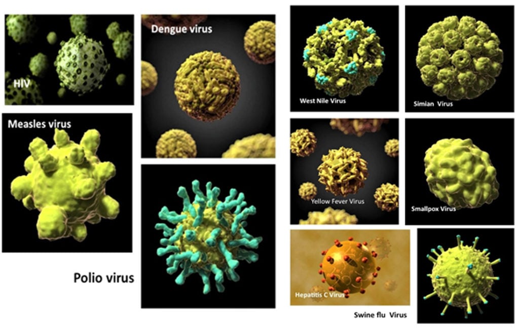
This out of this world image of coronavirus seems even more fascinating when one looks at the surface ultrastructure of many other deadly viruses. In Huffington Post, Jacqueline Howard wrote about the unique yet deadly beauty of viruses as seen in atomic resolution through cryoelectron microscopy with 3D reconstruction. See photos of these viruses in three-dimension reconstruction.
These 12 Viruses Look Beautiful Up Close But Would Kill You If They Could https://www.huffpost.com/entry/deadly-viruses-beautiful-photos_n_4545309
But, are they really that unique and alien-looking?
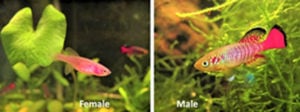
Adult male and female Nothobranchius guentheri, native to the island of Zanzibar, Tanzania. Males are about 5 cm in length while the colorless females are much smaller at 3.5 cm.
In a recent Science Blog article in Sulu Garden,I showed how the corona, the most salient external feature of the SARSCov virus, resembles the corona of the outer envelope of the embryo of the annual killifish. This rare fish belongs to a class of Cyprinodontid fish that live in temporary pools of fresh water in Africa and South America that dry up during the summer season. The population survives as embryos buried in the mud that hatches during the rainy season. They survive through a process called diapause wherein the embryo enters states of suspended animation, very similar to many instances found in insects.
Convergent evolution of the surface micro and nano structures in coronavirus and annual fish embryo. Thoughts while under “enhanced community quarantine” in Miag-ao (Iloilo Province, Panay Island, Philippines). http://sulugarden.com/articles/blog/science/
Despite the difference in size (annual fish embryo at 1 millimeter and coronavirus at approximately 120 nanometers), both the coronavirus and fish embryo share similar surface topology. Hence, I suggested that this might be considered an instance of convergent evolution.
“Convergent evolution is a process wherein two unrelated organisms independently develop the same traits or characteristics to address a similar environmental problem. An example of convergent evolution is the adaptations to flight. Different animals developed wings independently of each other to enable flight, such as bats, butterflies, pterosaurs and birds [https://www.sciencedaily.com/terms/convergent_evolution.htm]. The spikes that coronaviruses and annual fish embryos possess were developed independently to solve each of their own ecological situations. It is interesting that the annual fish embryo is 1 mm in diameter while the diameter of a coronavirus is 60 to 140 nm or about 1/100,000th of the fish embryo https://www.britannica.com/science/coronavirus-virus-group. So far away from each other as species goes, yet they created the same evolutionary solution to a similar problem. Although the coronavirus is technically not a life form, but simply a single strand of RNA (ribonucleic acid) particle, it does mutate often and is evolving faster than animals or plants do.”
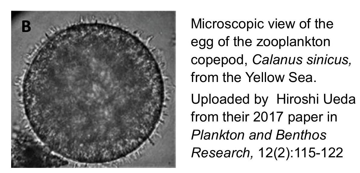 Other species of marine animals also show a diverse array of surface spikes, depending on the survival requirements. Some that need to attach to surfaces show spike patterns while others with different ecological issues show different morphology. For example, here is a photo of the zooplankton copepod that represents 80% of the zooplanktons in the Yellow Sea. It does show its own style of spikes on the surface of the outer egg envelope.
Pollen Grains
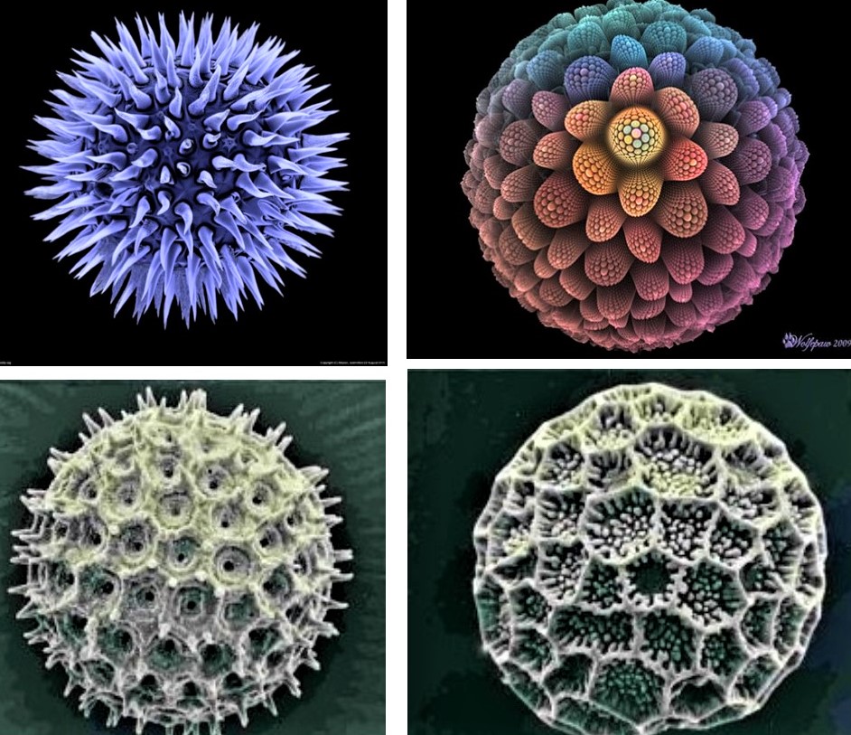
 This corona-like pattern is not only confined to marine and freshwater species. Perhaps the most striking diversity of surface structures can be found in pollen grains. A cursory search of pollen grain images in Pinterest and other websites demonstrates a vast array of surface modifications to allow pollen to attach to different substrates.
Here are some examples for you.
For those suffering from pollen allergy, the spikes make it possible for the pollen grains to attach to the nasopharyngeal passages and skin more efficiently.
Interested in seeing more? Here are the links to see these images:
https://www.pinterest.co.uk/pin/460141286903103442/
https://www.pinterest.co.uk/pin/460141286903103403/
https://www.pinterest.co.uk/pin/132363676521662748/
https://allthatsinteresting.com/objects-under-a-microscope/2
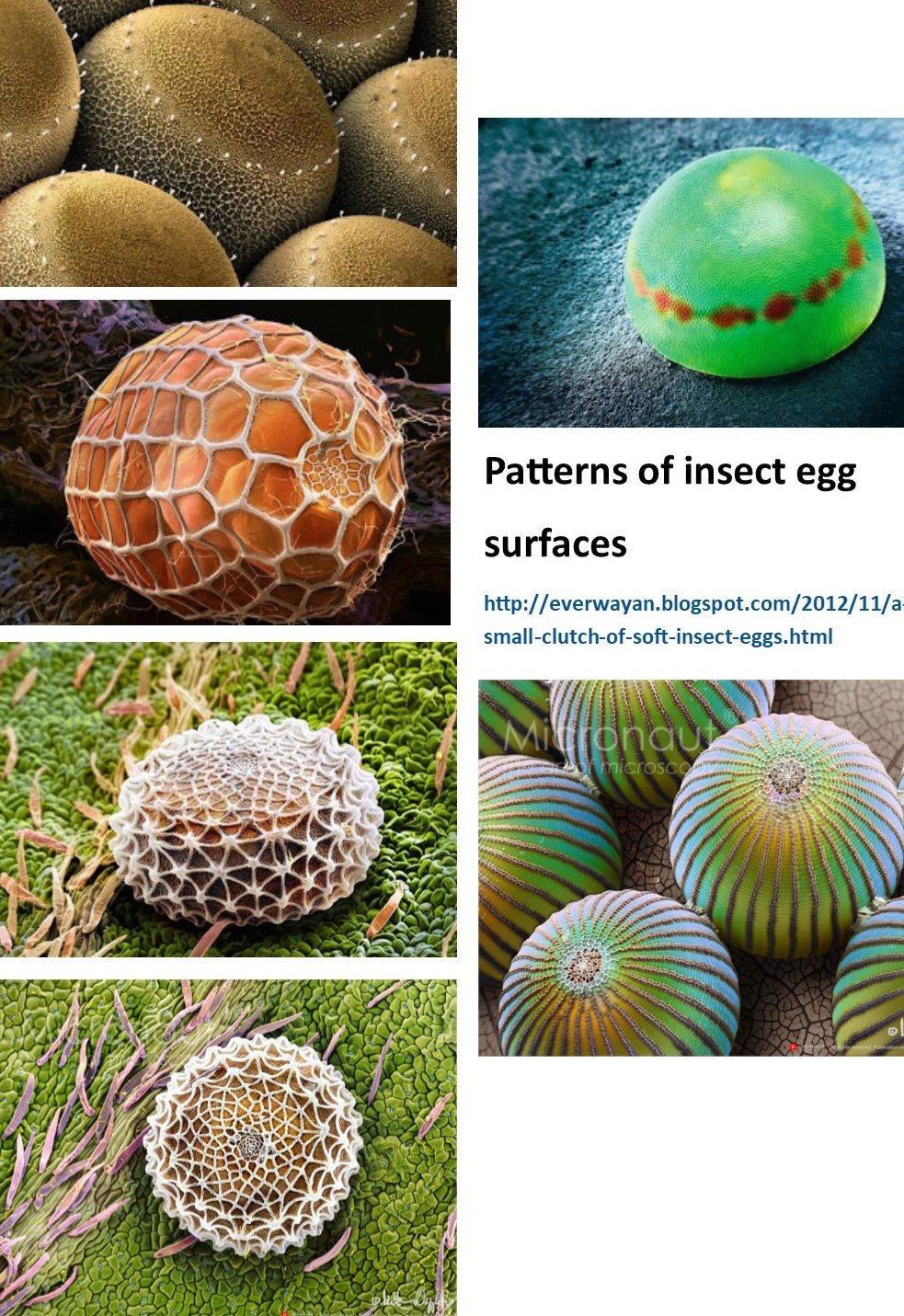
Surface structure of Insect eggs
Even insect eggs show a diverse, alien-like surface structure to attach to leaves of plants and other insects
Plants
The ‘bangkal tree’ is an example of diversity of structures that even higher plants demonstrate.
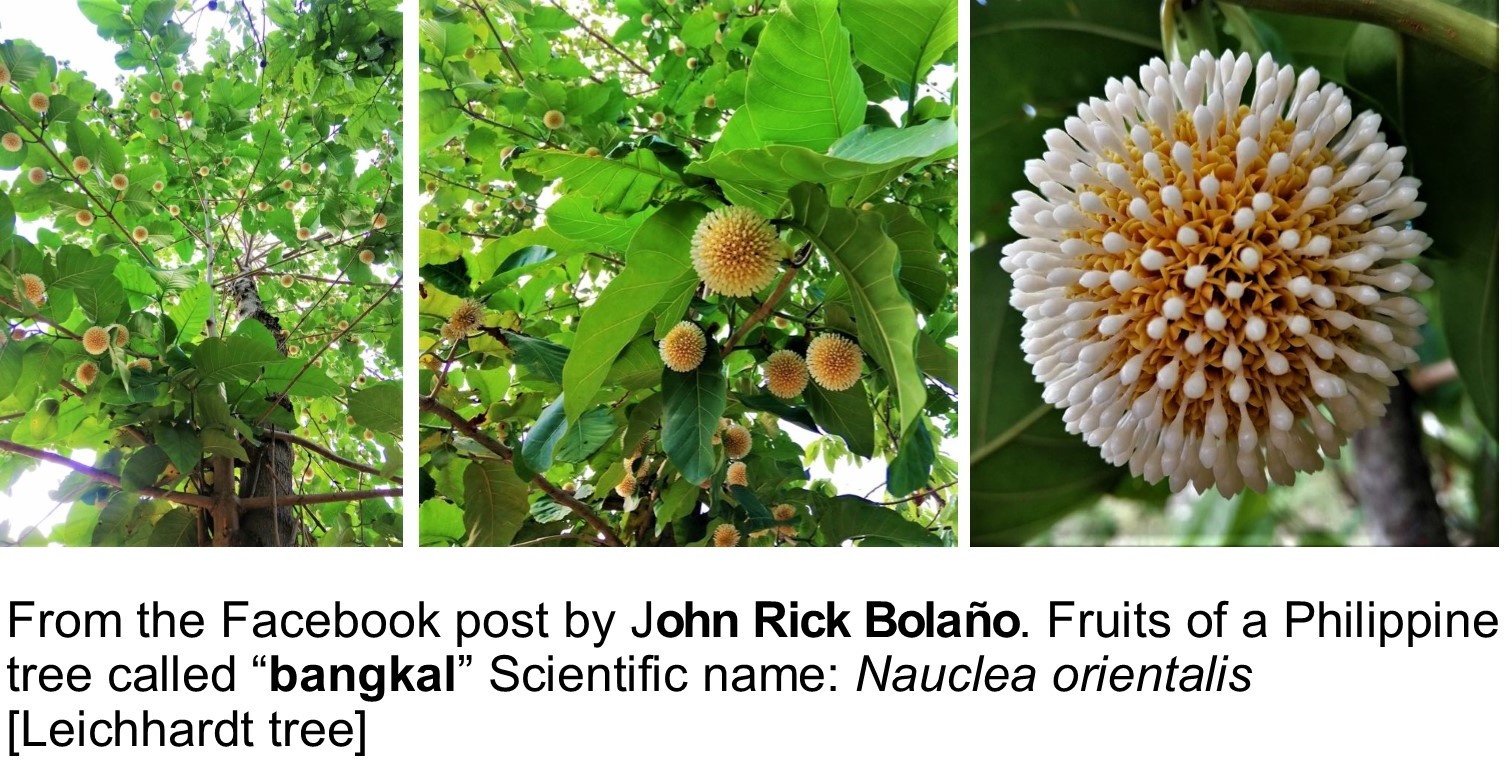 Though viruses seem out of this world, their surface morphology is more common in nature than we think. What is shown here is just a glimpse of how diverse these microscopic surface structures can be. I am sure one can find even better examples in the web.
The virus is an ancient entity whose origins remain in debate to this day. But everyone agrees that it likely predated or co-evolved very early in the evolution of life as we know it today and continued to mutate through each stages of evolution. https://en.wikipedia.org/wiki/Viral_evolution
Perhaps, the evolution of the intricate surface structures in seeds, pollen grains, fish eggs and even some plants might have originated from viruses?
Jonathan R. Matias
Poseidon Sciences R&D
Miag-ao, Iloilo 5023 Philippines
www.poseidonsciences.com
June 16, 2020
If you wish to comment, please send email to [email protected]

